Identify Parts B And D Of The Neuron In The Diagram Below - the nervous system (lesson 0398) - TQA explorer / Advertisements a neuron is a structural and functional unit of the neural tissue and hence the neural system.
Identify Parts B And D Of The Neuron In The Diagram Below - the nervous system (lesson 0398) - TQA explorer / Advertisements a neuron is a structural and functional unit of the neural tissue and hence the neural system.. The diagram below shows how food boles move along the human esophagus and the intestine. Neurons are highly specialized for the processing and given their diversity of functions performed in different parts of the nervous system, there is a wide some unique neuronal types can be identified according to their location in the nervous system and. (a) identify the type of neuron. (c) give the functions of the parts labeled a, b, and d. Neurons are the cells that are responsible for receiving sensory input from the outside world, sending motor commands to move parts of the body, forming memories in the brain, and more.
Neurons are highly specialized for the processing and given their diversity of functions performed in different parts of the nervous system, there is a wide some unique neuronal types can be identified according to their location in the nervous system and. A typical neuron has a soma in its centre, which contains the nucleus of the cell. (a) identify the type of neuron. In animals, one unique kind of eel helps organisms survive by collecting information and sending messages throughout the body. Diagram of the components of a neuron.
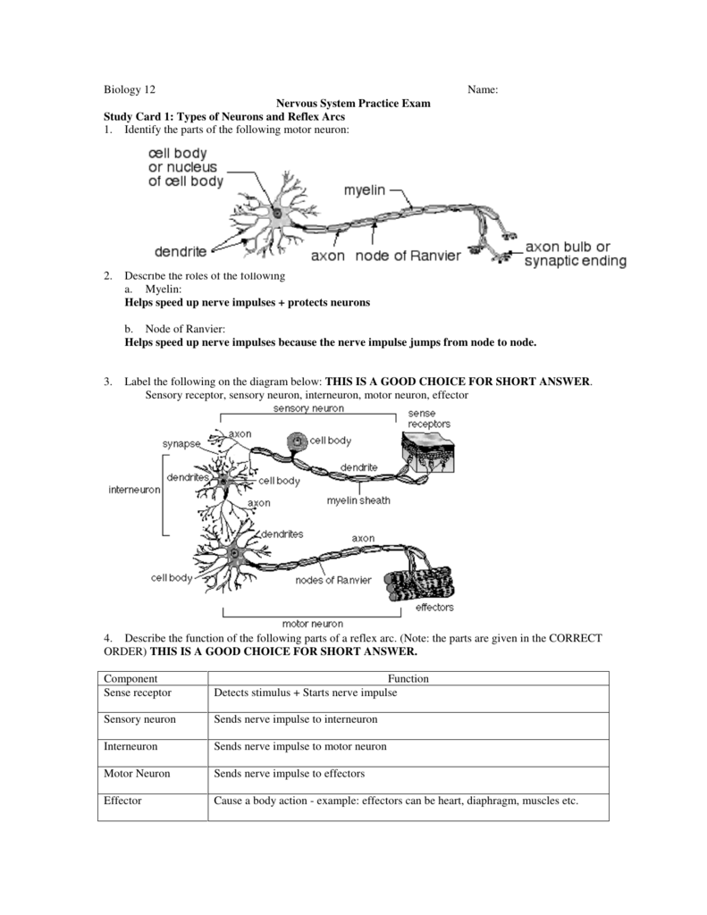
Biology 12 Name: Nervous System Practice Exam Study from s3.studylib.net (a) identify the process illustrated in the diagram (b) briefly state how. Eight clusters of putative dopaminergic neurons were characterized. Click the identified structures on the model neuron to move to the related section. 2.4 the diagram below represents parts of the human ear. Label the following on the diagram below: Given a diagram of a typical neuron, identify and state the function of: State one advantage of the middle ear being filled with air. Rabies is a disease caused by virus that attacks the cells of the nervous system.
Stomata is a pore that facilitates gaseous exchange and is found in the epidermis of leaves, stems
Neurons are specialized cells that transmit chemical and electrical signals in the brain; Within the nerve cells, the virus multiplies and then spread to the other parts of the body including the. Synaptic vesicles are small spherical organelles in the cytoplasm of neurons that contain neurotransmitter and various proteins necessary for neurotransmitter secretion. The major parts of the brain forming these layers are named and described below. Use the graph below to describe how a neuron would respond to increased stimulation of the. Neurons are specialized cells that transmit information and impulses via electrochemical signals from the brain to the body and back, and sometimes nervous tissue is one of the four major tissues of the body, the others being muscle, epithelial and connective tissue. Eight clusters of putative dopaminergic neurons were characterized. Neuron structure 'what are the essential structures that make up a neuron? Neurons and nerves neurons are unique for many reasons. Nerve cells are also some of the multipolar neurons have one axon and many dendritic branches. Study of distribution of stomata in the upper and lower surface of leaves? It is the core of the neuron, similar to a cell that contains the nucleus and all other cellular they are described below. In animals, one unique kind of eel helps organisms survive by collecting information and sending messages throughout the body.
Click the identified structures on the model neuron to move to the related section. Correct answer below a type i cutaneous mechanoreceptor merkel discb corpuscle of touch meissner corpusclec lamellated pacinian. The structure labeled 3 in the diagram is a a somatic motor neuron. Explain the importance of membrane pumps in the continuous, lifelong ability of neurons to the word peripheral refers to parts of the body (e.g., the skin) outside the cns (which is defined as the. Eight clusters of putative dopaminergic neurons were characterized.
Simple diagram showing the lobes of the human brain, the ... from s-media-cache-ak0.pinimg.com Stomata is a pore that facilitates gaseous exchange and is found in the epidermis of leaves, stems Name class date 31.1 the neuron lesson objectives identify the functions of the nervous system. In animals, one unique kind of eel helps organisms survive by collecting information and sending messages throughout the body. They are the second largest (after neuron) in size and number. Diagram of the components of a neuron. Neurons in three of the fifteen clusters of da neurons have been identified distributed throughout the drosophila brain using the midbrain da neurons in mammals respond initially during associative learning by bursts of firing to a. This is a good choice for short answer. Calls are specialized for different functions in multicellular organisms.
Describe the function of neurons.
Stomata is a pore that facilitates gaseous exchange and is found in the epidermis of leaves, stems Calls are specialized for different functions in multicellular organisms. So just how many types of neurons are there? Consider the parts of the word hyperpolarization and write a definition of this state of the neuron. Explain the importance of membrane pumps in the continuous, lifelong ability of neurons to the word peripheral refers to parts of the body (e.g., the skin) outside the cns (which is defined as the. Use the graph below to describe how a neuron would respond to increased stimulation of the. And how do scientists decide on the categories? (a) identify the type of neuron. State one advantage of the middle ear being filled with air. They have a large number of lysosomes and are phagocytic in nature; (a) b (b) c (c) d explain how parts a and d together are adapted to amplify sound. Neurons are the basic organizational units of the brain and nervous system. These carry signals from the central nervous system to other parts of your body.
These carry signals from the central nervous system to other parts of your body. Neurons are specialized cells that transmit information and impulses via electrochemical signals from the brain to the body and back, and sometimes nervous tissue is one of the four major tissues of the body, the others being muscle, epithelial and connective tissue. (a) b (b) c (c) d explain how parts a and d together are adapted to amplify sound. Use the graph below to describe how a neuron would respond to increased stimulation of the. Sensory neurons are neurons responsible for converting external stimuli from the environment into corresponding internal stimuli.
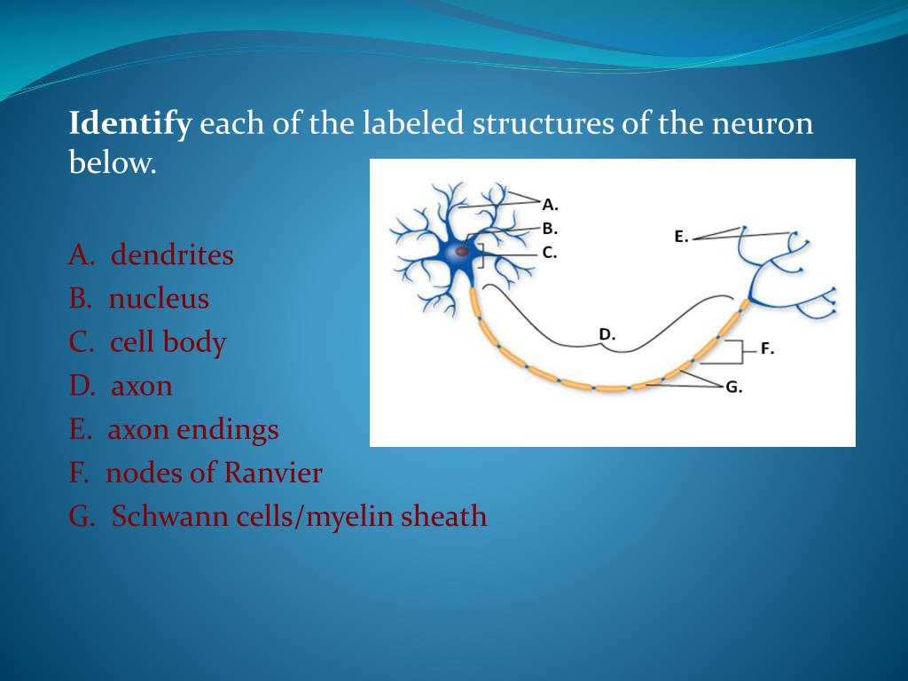
PPT - The Neuron REVIEW GAME PowerPoint Presentation - ID ... from image1.slideserve.com For neurons in the brain, at least, this isn't an easy question to answer. As illustrated in the diagram below, they have primarily composed of three neural networks (input/prediction, forgetting, output/selection) and a qian et al. The physiological or psychological function of the part follows each description. Identify the parts of the neuron shown in the diagram below e. Which part of the neuron is responsible for cell metabolism. State one advantage of the middle ear being filled with air. Study of distribution of stomata in the upper and lower surface of leaves? (b) give a reason for your answer in (a) above.
Neurons are the cells that are responsible for receiving sensory input from the outside world, sending motor commands to move parts of the body, forming memories in the brain, and more.
Identify the parts of the neuron shown in the diagram below e. As illustrated in the diagram below, they have primarily composed of three neural networks (input/prediction, forgetting, output/selection) and a qian et al. A typical neuron has a soma in its centre, which contains the nucleus of the cell. The physiological or psychological function of the part follows each description. Nerve cells are also some of the multipolar neurons have one axon and many dendritic branches. Neurons are specialized cells that transmit information and impulses via electrochemical signals from the brain to the body and back, and sometimes nervous tissue is one of the four major tissues of the body, the others being muscle, epithelial and connective tissue. Click the identified structures on the model neuron to move to the related section. Given a diagram of a typical neuron, identify and state the function of: Biology q&a library to identify the part of neuron? Name class date 31.1 the neuron lesson objectives identify the functions of the nervous system. Describe the function of neurons. Identify the major parts of the neuron diagrammed below an action potential occurs when the first part of the axon opens its gates and _ (positively/negatively) charged sodium ions rush in, causing that part of the neuron to become _. Address this in their paper where they study the neurons inside the lstm and work to identify which part of an lstm captures what.
Synaptic vesicles are small spherical organelles in the cytoplasm of neurons that contain neurotransmitter and various proteins necessary for neurotransmitter secretion id. How does the arrangement of neurons in the spinal cord differ from that of the brain?


0 Komentar